We are specialised in visualising the cell and its molecular constituents, aiming for clarity and insight besides producing stunning animated artwork. Our work is regarded as quintessential in its field; we aim to produce cutting-edge visualisations, serving biomedical education and research institutions, in addition to biotechnology and pharmaceutical companies.
Our scientific background, trained as molecular biologists to the PhD level, visualisation skills and experience, has allowed us to assist our clients in effectively and faithfully conveying their findings to their many audiences.
We look forward to working closely with our clients, continuing to raise the bar for the visualisation and communication of molecular and cellular research findings.

Solutions
At Cellscape we are very enthusiastic about visualising the intricate functions of the cell and its molecular machinery. We provide our clients with effective solutions to depict their biomedical findings and insights based on our considerable experience gained from projects with leading scientists including two Nobel Laureates.
Our approach
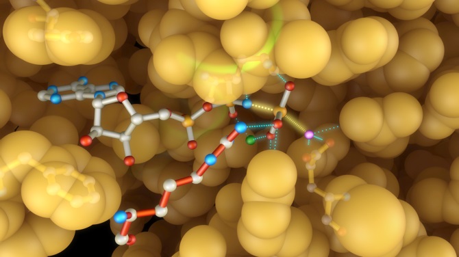
We use a variety of generic and specialised molecular modelling and visualisation methods, together with high-end computer graphics modelling and animation methods. We are also developing an open-ended framework that is intended to enable us and our clients to develop exceedingly more complex and accurate molecular models that span the nano to the whole cell scale and beyond.
Outcome
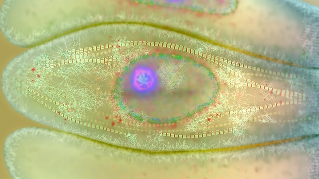
We have established a myriad of effective solutions for modelling and visualising the actions and interactions of molecules and
molecular assemblies at multiple spatial scales. These have allowed us to produce spectacular and lucid molecular and subcellular
behaviours, achieved by developing compelling mechanistic and visualisation solutions for a wide range of molecular and
subcellular phenomena.
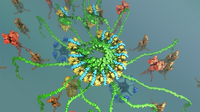
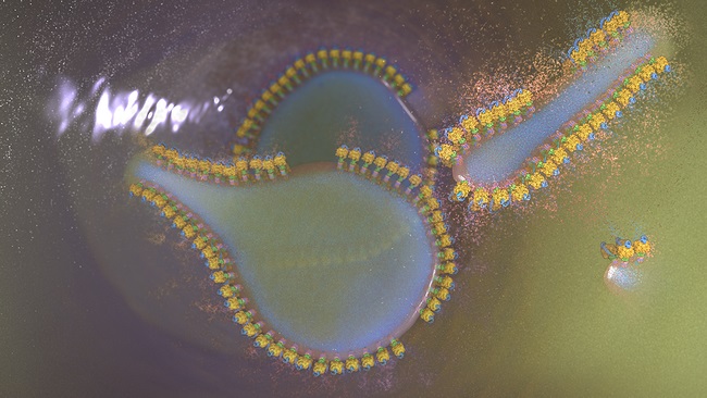
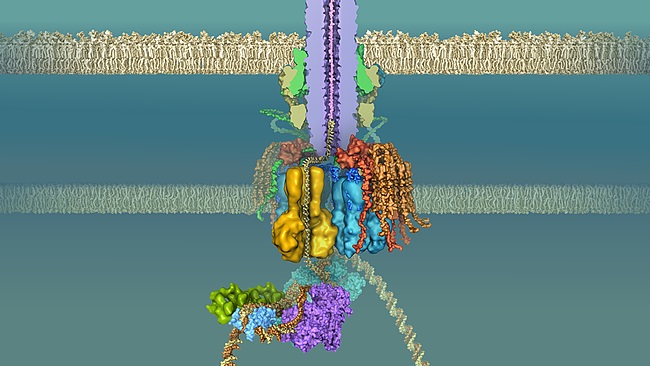
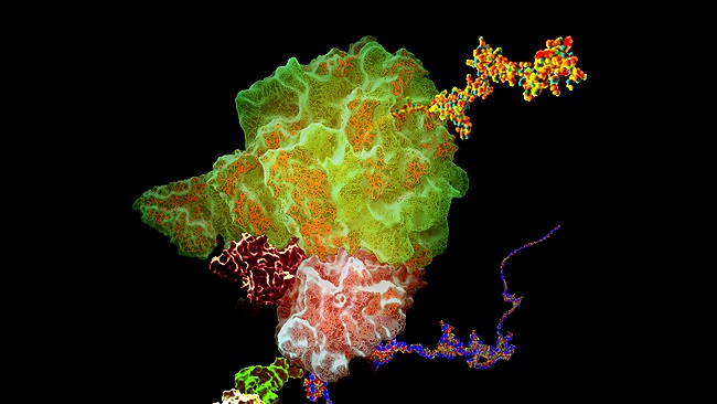
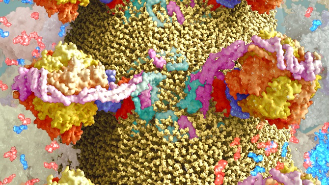
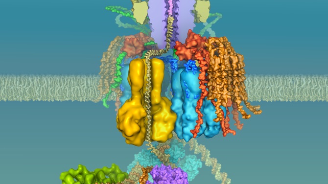
For example, we have developed effective solutions to visualise several ribosomal structures, from bacteria
to man; these were determined by several methods, requiring an in-depth understanding of the subject and the careful
handling and integration of data from the different methods and species used.
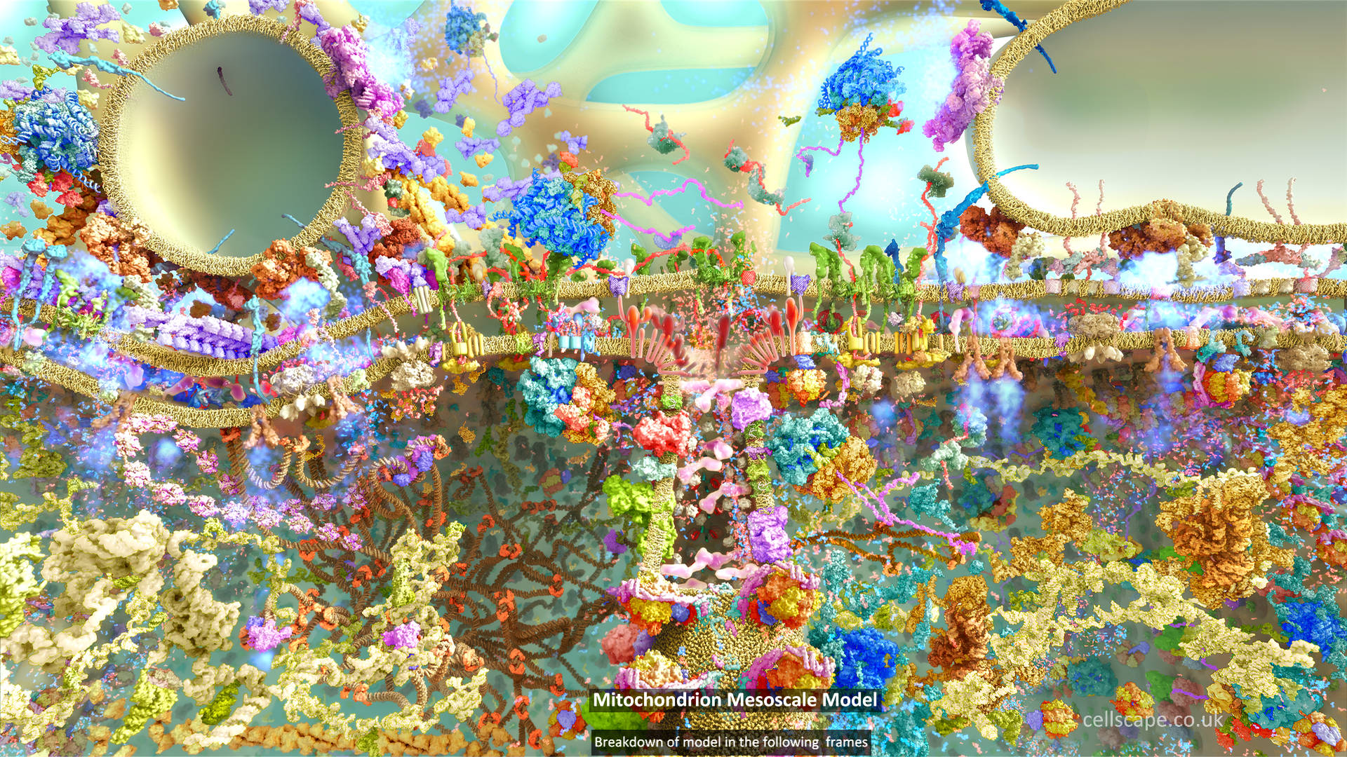
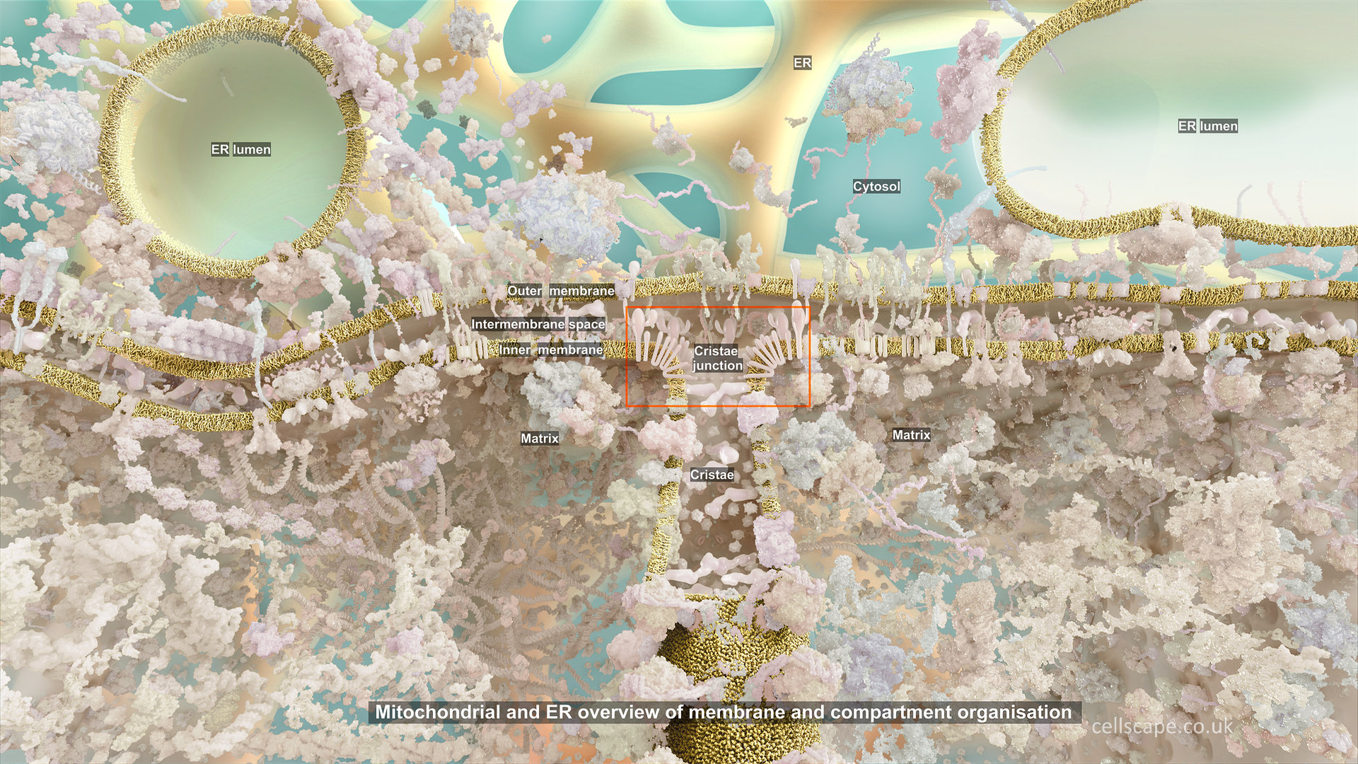
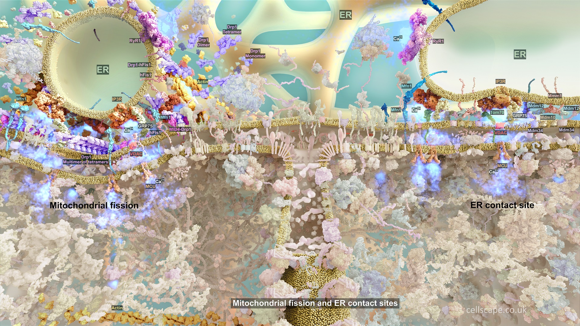
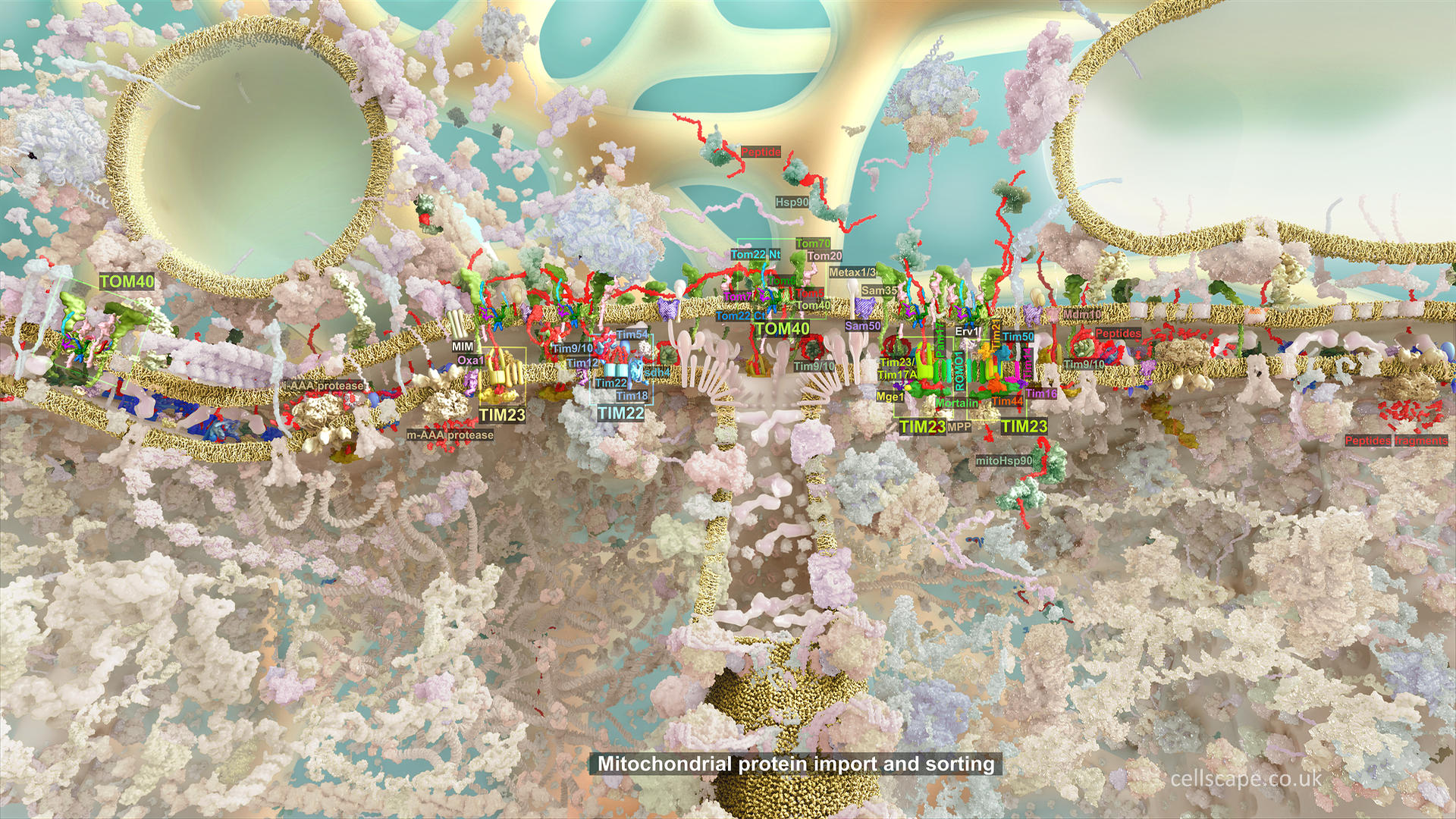
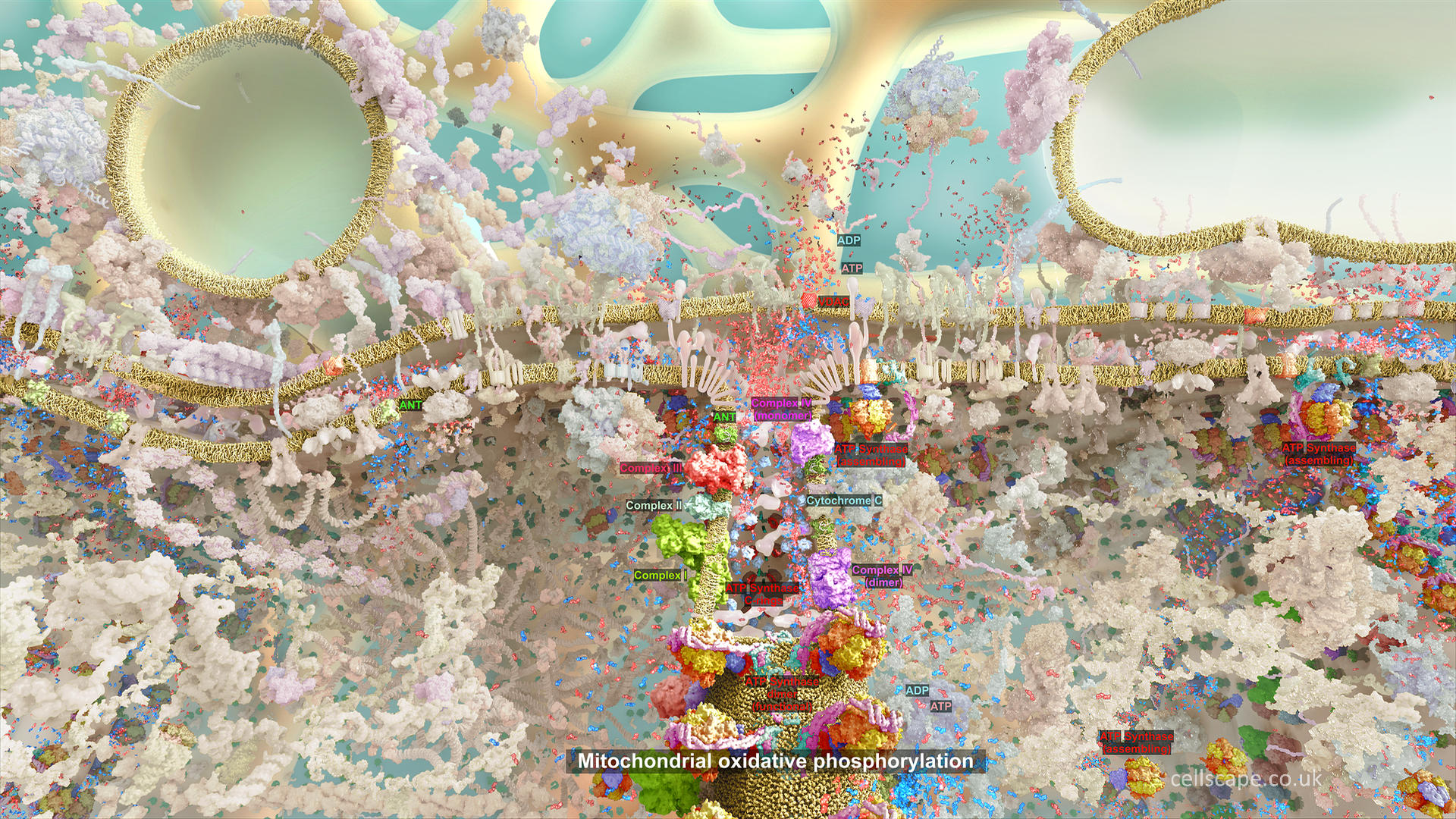
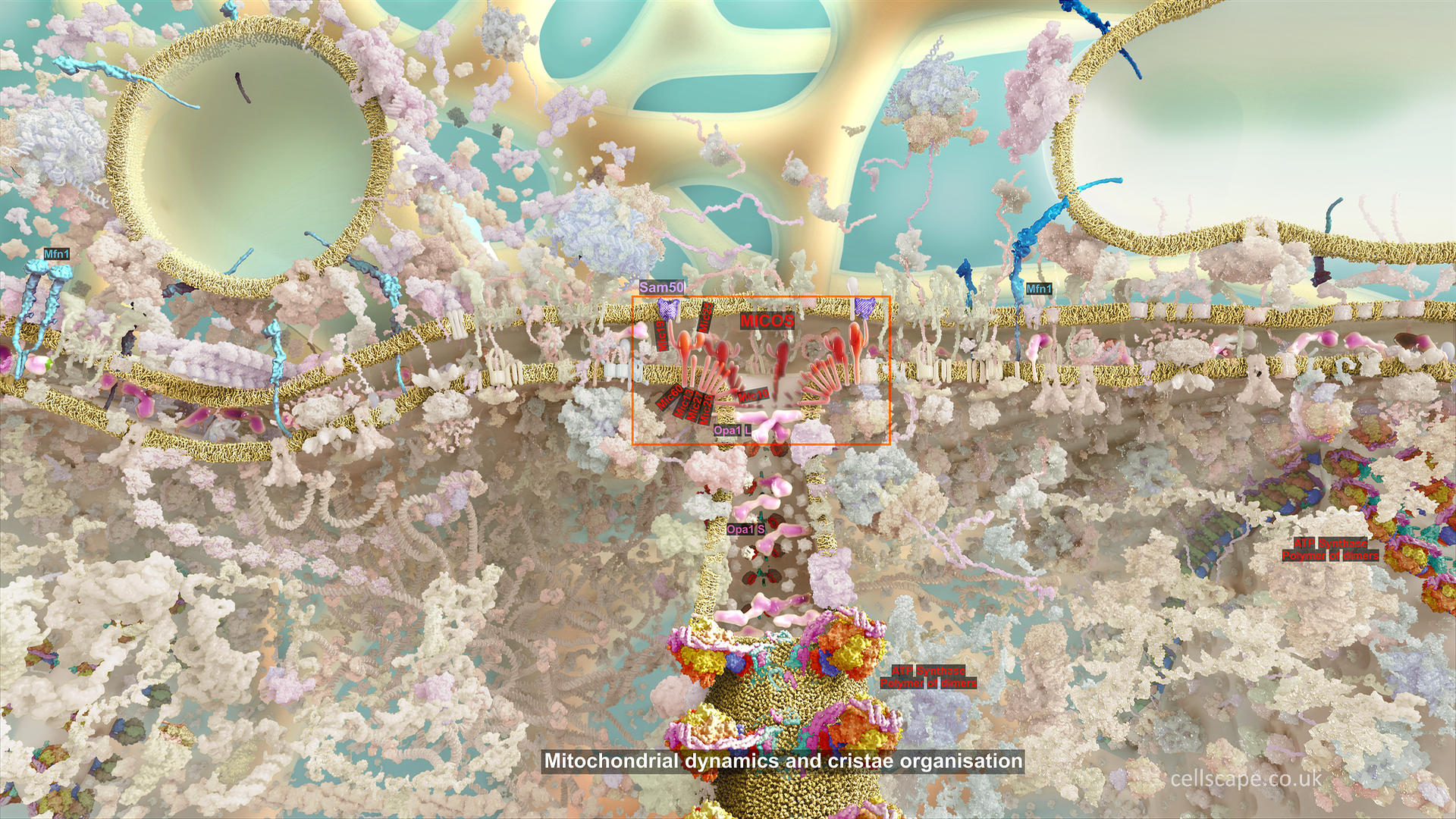
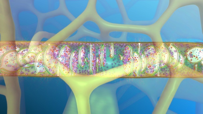
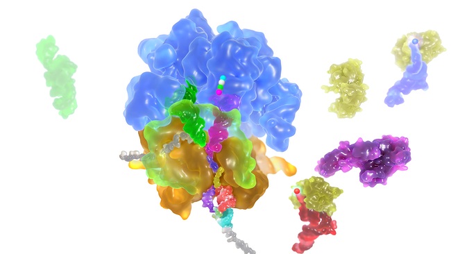
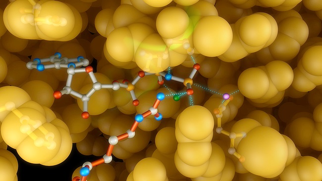
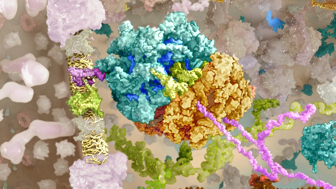
Another example of our work is a detailed mitochondrion mesoscale model which is based on approximately 45 pdb and
15 EMD structures, 20 3D models of proteins of unknown structure, 11 models of proteins of partially known structure
and several models for nucleic acids (e.g. the nucleoid DNA is 19600bp).
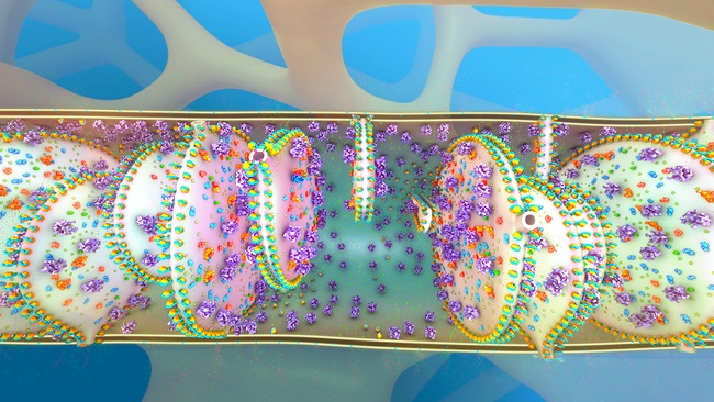
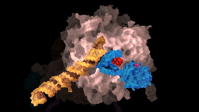
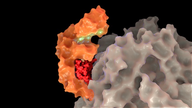
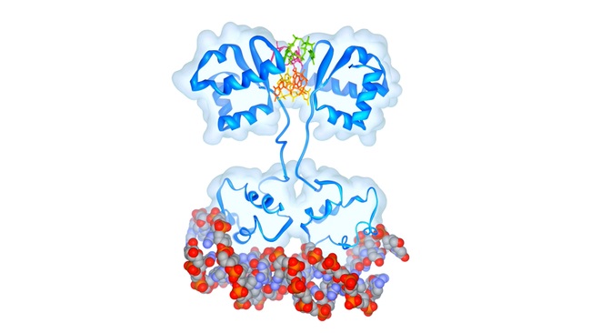
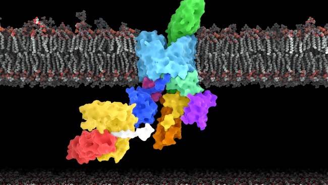
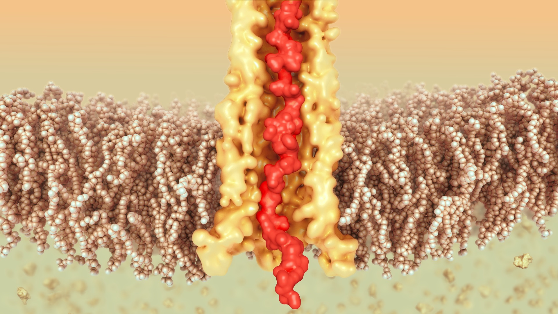
We depict innovatively the molecular machines of the cell, transforming them effectively and vividly from static models into
dynamic, functioning molecules, with consideration given to their known quantity, spatial distribution and their prolific
interactions and behaviour within their subcellular environments.
Biological context
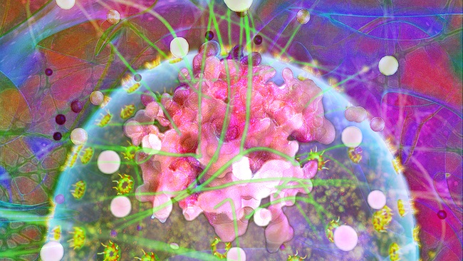
The cellular mesoscale to the microscale is the scale at which the complex characteristics of life begin to emerge. This scale contains a mix of self-organising molecular machinery, processes and circuits; an arena where the workings of the cell's constituents can be depicted to provide clarification of life phenomena when in a healthy or pathological state. Our outstanding visual work on molecular machines and the development of our meso-microscale models should empower us to assist our scientific and pharmaceutical clients in communicating the functioning of their molecular machines or their drug mechanisms of action in a biological context and, therefore, achieve a broader comprehension of the implications of their findings and products.
Clients